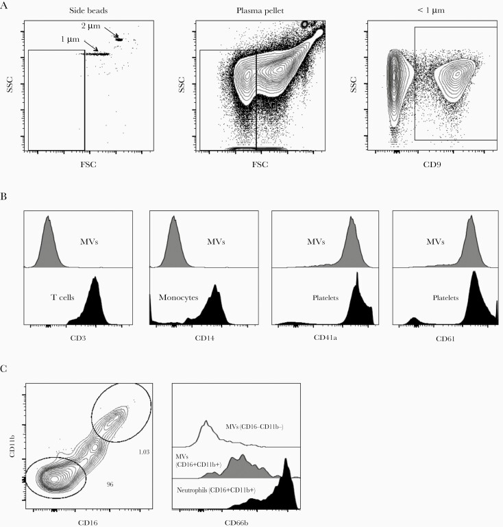
Figure 1.
Gating strategy to characterize microvesicles (MVs) by flow cytometry. (a) Microvesicles less than 1 μM were gated using size reference beads and CD9 expression. Representative contour plots showing side scatter (SSC) and forward scatter (FSC) or CD9 labeling of MVs from antiretroviral therapy-exposed human immunodeficiency virus-infected donors (n = 30); (b) CD3 staining on plasma-derived MVs or peripheral blood-derived T cells; CD14 staining on plasma-derived MVs or monocytes, and CD41a and CD61 staining on plasma-derived MVs or platelets; (c) coexpression of CD11b and CD16 on MVs and CD66b staining on plasma-derived MVs subsets or peripheral blood-derived neutrophils.

