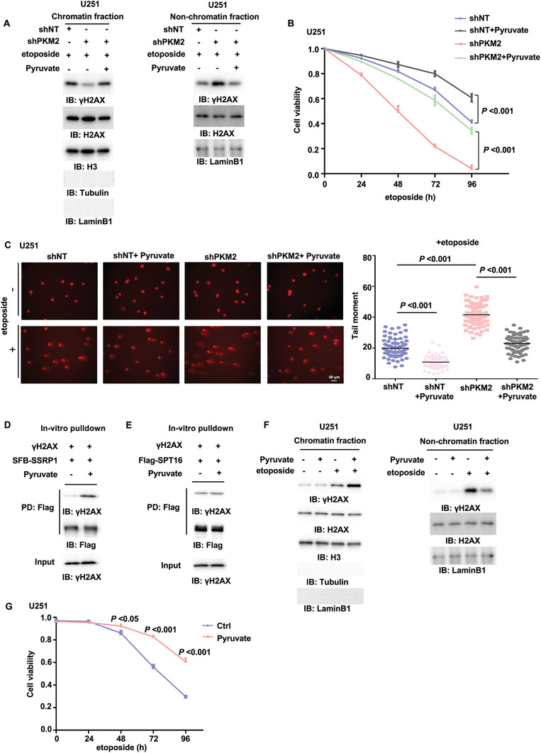
Figure 3.

Pyruvate increases the interaction between FACT and γH2AX, γH2AX levels in chromatin and tumor cell survival upon DNA damage. IP and IB analyses were performed with indicated antibodies. Data are representative of at least three independent experiments. A) U251 cells stably expressing shNT or shPKM2 were supplemented with or without pyruvate (10 × 10–3 m, 3 h) and then treated with etoposide (200 × 10–6 m, 1 h). Chromatin fraction (left panel) and nonchromatin‐bound fraction (right panel) were prepared. B) U251 cells stably expressing shNT or shPKM2 were supplemented with or without pyruvate (10 × 10–3 m, 3 h) and then treated with etoposide (200 × 10–6 m) for indicated time. Cell viability was determined. Data represent the mean ± SD of the viability of the cells from three independent experiments (two‐tailed Student's t‐test). C) U251 cells stably expressing shNT or shPKM2 were supplemented with or without pyruvate (10 × 10–3 m, 3 h) and then treated with or without etoposide (500 × 10–6 m) for 3 h. The representative images of the comet assay in these cells were shown (left panel). Scatter dot plot (right panel) represents the tail moments in the comet assay from cells treated with etoposide, expressing shNT with (n = 75) or without (n = 83) pyruvate, or cells expressing shPKM2 with (n = 78) or without (n = 78) pyruvate. Data represent mean ± SD of the tail moments (Mann Whitney test). Data are representative of three independent experiments. D,E) SSRP1 or SPT16 was immunoprecipitated and purified from U251 cells stably expressing D) SFB‐SSRP1 or E) Flag‐SPT16. γH2AX was immunoprecipitated and purified from etoposide‐treated U251 cells. The in vitro pulldown experiment was performed by incubating the purified SSRP1 or SPT16 with purified γH2AX in the absence or presence of pyruvate (1 × 10⁻³ m). PD: pulldown. F) U251 cells were supplemented with pyruvate (10 × 10–3 m, 3 h) and then treated with etoposide (200 × 10–6 m, 1 h). Chromatin fraction (left panel) and nonchromatin‐bound fraction (right panel) were prepared. G) U251 cells were supplemented with or without pyruvate (10 × 10–3 m, 3 h) and then treated with etoposide (200 × 10–6 m) for indicated time. Cell viability was determined using Trypan blue staining. Data represent the mean ± SD of the viability of the cells from three independent experiments (two‐tailed Student's t‐test).
