
Figure 3.
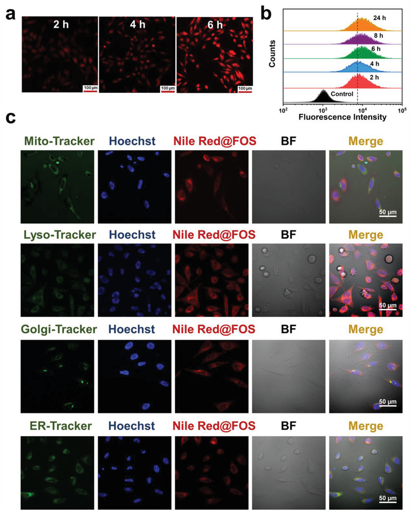
a) CLSM images of HeLa cells incubated with Nile Red@FOS for various periods (scale bar: 100 µm). b) Cell uptake of Nile Red@FOS with different incubation periods by flow cytometry analysis. c) High‐magnification CLSM images of HeLa cells incubated with Nile Red@FOS for 12 h. Mitochondria (Mito), lysosomes (Lyso), Golgi apparatus (Golgi), and endoplasmic reticulum (ER) were stained with the corresponding green fluorescent tracker. The nucleus was marked blue by the classic staining reagent Hoechst 33342 (Hoechst). The bright field is abbreviated as BF.