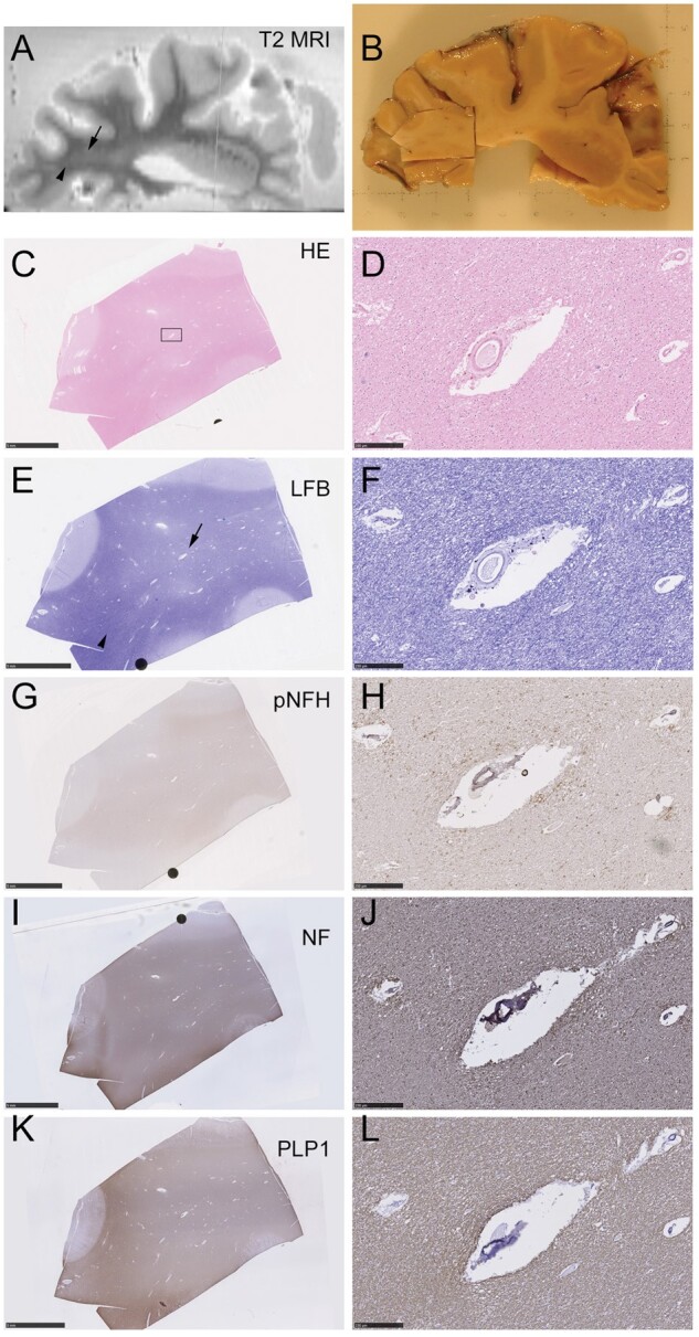
FIGURE 4.

Ex vivo MRI-directed sampling of T2 hyperintense subcortical white matter. RADC-derived tissue stained for hematoxylin and eosin (H&E) and Luxol fast blue (LFB) and immunolabeled for pNfH, pan-neurofilament (NF), and myelin (PLP1). (A) Ex vivo T2-weighted MRI of a 1-cm-thick slice of fixed brain tissue. A region of WMH is visible (arrow), adjacent to an area of nonhyperintense normal-appearing white matter (arrowhead). (B) At gross pathology, the sampled tissue block contains the region of WMH seen in panel (A). (C, D) H&E staining of the sampled tissue block is shown in panel (B). The low magnification view in panel (C) shows the region (rectangle) that is magnified in the right-hand panels, containing a landmark blood vessel. (E, F) LFB-stained section exhibits white matter pallor (arrow) in region identified as WMH in the T2-weighted scan in panel (A). This contrasts with the more densely stained area (arrowhead) identified as normal appearing white matter in panel (A). (G, H) Immunolabeling for pNfH (SMI35 antibody) is not noticeably more pronounced in the area of WMH, though high magnification confirms the vasculocentric axonal pattern (H). (I, J) Axonal neurofilament (pan-neurofilament antibody) shows no marked loss of positivity in the WMH region. (K, L) Myelin immunolabeling (PLP1 antibody) resembles LFB in terms of pallor within the WMH region (K). Scale bars: 5 mm in low-magnification images, 250 μm in high-magnification images.
