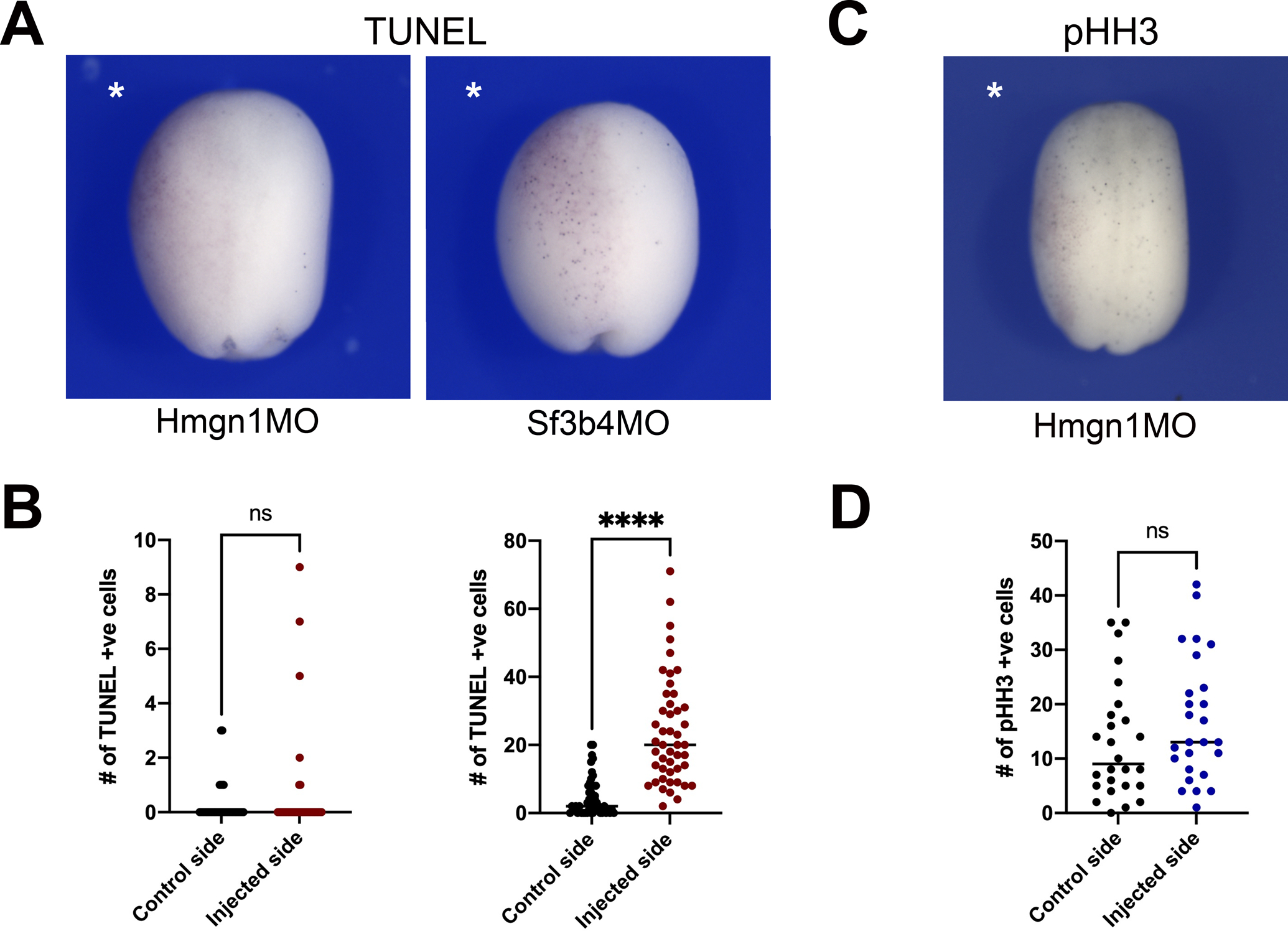
Figure 5: Hmgn1 knockdown does not affect cell death or proliferation in the ectoderm.

(A) TUNEL staining of representative Hmgn1 and Sf3b4 morphant embryos at stage 15. The injected side is indicated by an asterisk. Dorsal view, anterior to top. (B) Quantification of the number of TUNEL-positive cells in control and injected sides of Hmgn1MO (n=40; left graph) and Sf3b4MO (n=47; right graph) injected embryos at stage15. (C) pHH3 immunostaining of a representative Hmgn1 morphant embryos at stage 15. (D) Quantification of the number of pHH3-positive cells in control and injected sides of Hmgn1MO injected embryos (n=26). (B, D) Each dot represents one embryo. p-values were calculated using unpaired t-test with Welch’s correction, **** p<0.0001; ns: not significant.
