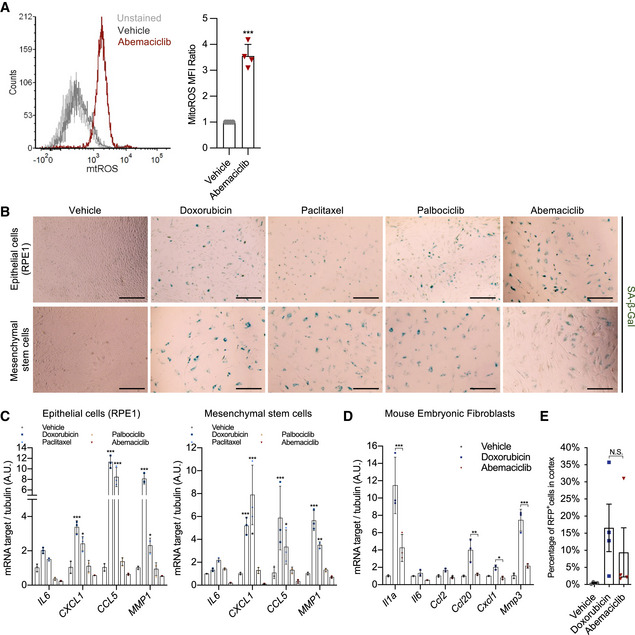
Figure EV4. CDK4/6i induces cellular senescence without pro‐inflammatory SASP.

- BJ cells were treated with vehicle or abemaciclib and stained for mitochondria ROS. Relative values of ROS level were plotted (n = 4 independent experiments).
- Human hTERT‐RPE1 and lung mesenchymal stem cells were treated with vehicle (DMSO for 8 times in 24 h) or doxorubicin (250 nM for 24 h) or paclitaxel (50 nM for 24 h) or palbociclib (1 μM for 8 times in 24 h) or abemaciclib (1 μM for 8 times in 24 h). At 8 dpt, treated cells were fixed and stained for SA‐β‐gal (scale bar, 1 mm; n = 3 independent experiments).
- At 8 dpt, RNA was isolated from treated cells and indicated NF‐κB‐associated SASP (NASP) genes were quantified by qRT–PCR relative to tubulin (n = 3 independent experiments).
- qRT–PCR of indicated genes was performed using mouse embryonic fibroblast (MEFs) 8 days after the indicated treatments (n = 3 independent experiments).
- RFP+ cells were sorted from renal cortex of doxorubicin‐ or abemaciclib‐treated p16‐3MR mice. The percentage of RFP+ cells in total cells was plotted (n = 4 mice/group).
Data information: Data are means ± SD. Two‐way ANOVA (C and D). One‐way ANOVA (E). *P < 0.05, **P < 0.01, and ***P < 0.001, N.S. = not significant.
