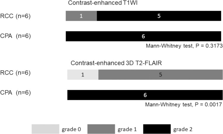
Fig. 1.
Distribution of the wall enhancement grade of a RCC and a CPA on contrast-enhanced T1W- and 3D T2-FLAIR images.
Grade 2: most of the cyst wall and/or cyst inner margin enhanced; grade 1, some of the cyst wall and/or cyst inner margin enhanced; grade 0, no enhancement in the cyst wall and cyst inner margin. 3D T2-FLAIR, 3D T2 fluid-attenuated inversion-recovery; CPA, cystic pituitary adenoma; RCC, Rathke’s cleft cyst; T1W, T1-weighted.

