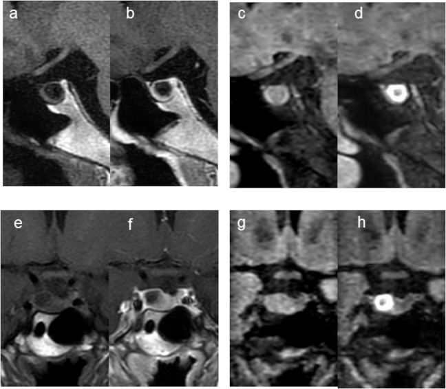
Fig. 2.
A 39-year-old woman with CPA (Case 8). Compared with the pre-contrast sagittal and coronal T1W images (a and e), the post-contrast sagittal and coronal T1W images (b and f) show cyst wall enhancement (grade 2). Pre- and post-contrast 3D T2-FLAIR sagittal (c and d) and coronal images (g and h). The post-contrast sagittal and coronal images (d and h) show remarkable, donut-like enhancement along the inner margin of the cyst (grade 2). The 3rd observer judged this lesion as an equivocal CPA (scale 3) at the 1st session but changed the confidence level to scale 5 at the 2nd session. 3D T2-FLAIR, 3D T2 fluid-attenuated inversion-recovery; CPA, cystic pituitary adenoma; T1W, T1-weighted.

