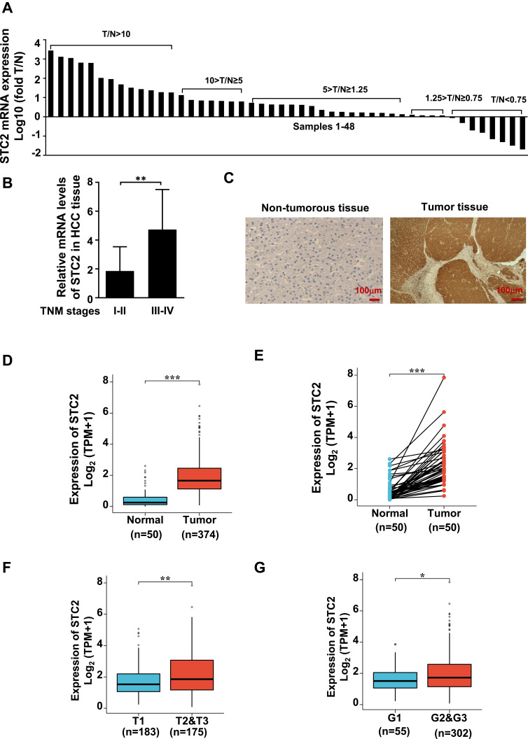
Figure 1.
STC2 was highly expressed in HCC and correlated with HCC progression. (A) STC2 mRNA levels were determined by qRT-PCR and compared between tumor (T) and adjacent non-tumorous tissues (N). The ratio of T/N was used as a measure of increased (T/N ≥ 1.25), decreased (T/N < 0.75), or no changed expression of STC2 (1.25 > T/N ≥ 0.75). (B) The mRNA levels of STC2 were compared between TNM stages III–IV and stages I–II. (C) STC2 protein in tumor and non-tumorous liver tissues was stained with immunohistochemistry with anti-STC2 antibody (1:100). (D) TCGA-LIHC database was used to explore the difference of STC2 mRNA between HCC tumor tissues (n=374) and normal tissues (n=50). (E) STC2 mRNA expression levels in HCC tumor tissues were compared to their corresponding non-tumorous tissues (n=50) based on TCGA-LIHC cohort. (F and G) Relationship analysis of STC2 mRNA expression levels with tumor stages and differentiated degrees of HCC on TCGA-LIHC tissue samples. *p<0.05; **p<0.01; ***p<0.001.

