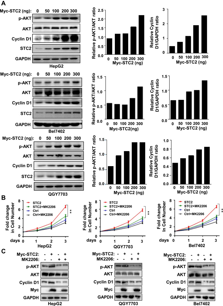
Figure 3.
STC2 promoted HCC cell growth by activation of AKT. (A) HepG2, Bel7402 and QGY7703 were transiently transfected with increasing concentrations of Myc-STC2 plasmid. After 24 h of transfection, cell lysates were analyzed for expression of STC2, AKT and its phosphorylated form, Cyclin D1. The expression of GAPDH was served as a loading control. The protein expression was determined by Western blotting. (B and C) HepG2, Bel7402 and QGY7703 were transiently transfected with Myc-STC2 plasmid and treatment with AKT inhibitor MK2206 (1 μM). The cell growth was detected with MTT (B). The cell lysates were analyzed for expression of STC2, Cyclin D1, AKT and its phosphorylated form (C). **p<0.01 (vs respective controls).

