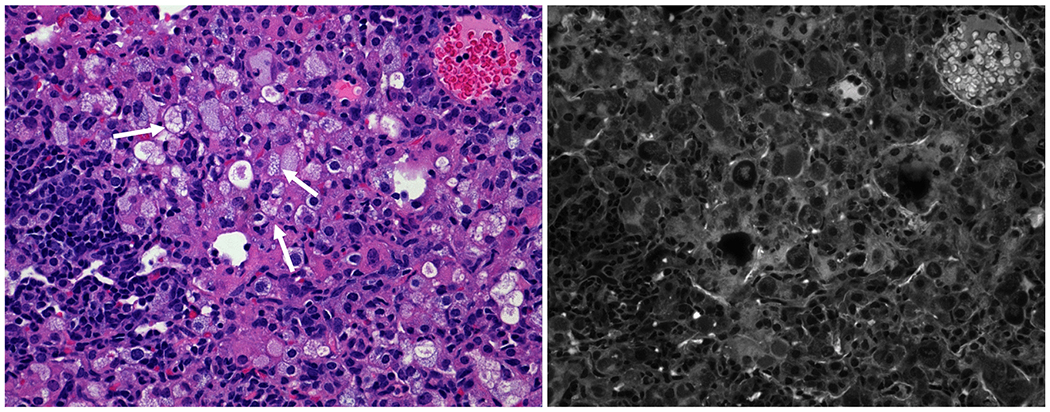
Fig. 6. Accumulation of ofloxacin signal in activated macrophages in lactoferrin treated Mtb infected mice.

H&E staining of inflammatory foci within rHLF therapeutically treated mouse lung revealed acute regions of highly activated “foamy” macrophage-phenotypic cells (white arrows). Mutispectral imaging correlates the presence of fluorescence which overlaps presence of activated cells, located within the focal granulomatous regions. Representative section, 100× magnification.
