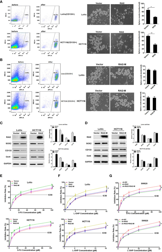
Figure 5.
RAI2 inhibited CRC stem cell-like properties and increased the chemosensitivity of CRC cells to 5-FU and L-OHP. (A) Left panel: Representative images showing CD133-positive cell proportion before and after fluorescence-activating cell sorting by CD133 antibody in LoVo/HCT116 cells. Right panel: Representative images showing sphere-forming ability affected by RAI2 re-expression in LoVo/HCT116 cells. (B) Left panel: Representative images showing CD133-positive cell proportion before and after fluorescence-activating cell sorting by CD133 antibody in LoVo/HCT116 cells. Right panel: Representative images showing sphere-forming ability affected by RAI2-M re-expression in LoVo/HCT116 cells. (C) Western blot of CD133, SOX2 and OCT4 expression in RAI2 re-expressed or vector control cells. (D) Western blot of CD133, SOX2 and OCT4 expression in RAI2-M re-expressed or vector control cells. (E) Representative curves of growth inhibitory effects of 5-FU in LoVo/HCT116 cells with or without re-expression of RAI2/RAI2-M in different concentrations (0.00, 6.25, 12.50, 25.00, 50.00, 100.00μM). (F) Representative curves of growth inhibitory effects of L-OHP in LoVo/HCT116 cells with or without re-expression of RAI2/RAI2M in different concentrations (0.00, 1.00, 2.00, 4.00, 8.00, 16.00μM). (G) Growth inhibitory effect of 5-FU/L-OHP in SW620 cells with or without RAI2 knocked down and SW620 cells with both shRAI2 and RAI2-M transfected. The treatment concentrations of 5-FU in SW620 cells were (0.00, 6.25, 12.50, 25.00, 50.00, 100.00μM), and the treatment concentrations of L-OHP were (0.00, 2.00, 4.00, 8.00, 16.00, 32.00μM). The viability of cells was measured by MTT assay after 5-FU/L-OHP treatment for 48 hrs. p-values: *≤0.05; **<0.01.

