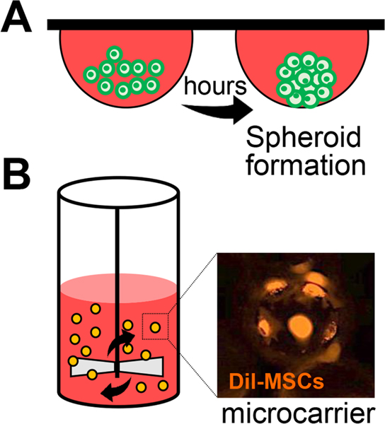
Figure 11.
Three-dimensional (3D) culture methods of MSCs.
A. Hanging drop culture for 3D spheroid formation. MSCs aggregate at the bottom tip of the drop and form a spheroid.
B. Spinner flask bioreactor with microcarriers. MSCs are attached to the surface of the microcarrier and grow as monolayers on the surface of a small sphere. Representative photograph of fluorescence (Dil)-labeled MSCs on microcarriers (Corning) at 4 hours after seeding.

