
Figure 1.
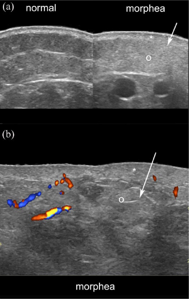
Ultrasound signs of active morphea. (a) Greyscale ultrasound (side-by-side comparison of normal perilesional with the lesional site) and (b) color Doppler present dermal thickening (*), loss of definition of the dermal and hypodermal border (arrows), increased echogenicity of the hypodermis (o) and increased dermal and hypodermal vascularity (b, in colors).