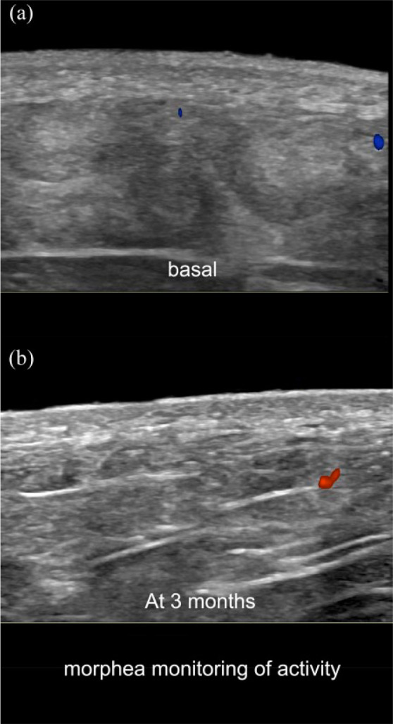
Figure 7.

Color Doppler ultrasound monitoring of morphea (transverse views; left gluteal region). (a) Basal examination with signs of activity (loss of the dermal and hypodermal border and increased hypodermal echogenicity). (b) Follow-up ultrasound at 3 months shows improvement in the definition of the border and decrease of the abnormal echogenicity of the hypodermis. Note the hypoechogenicity of the dermis in both images which is more prominent in the basal examination.
