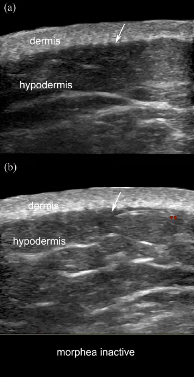
Figure 8.

Inactive morphea. (a) Greyscale and (b) color Doppler (transverse views; right arm) show a slight dermal thickening with decreased dermal echogenicity. Note that the dermal-hypodermal border is well-defined and there are no signs of increased echogenicity of the hypodermis or hypervascularity (arrows). Prominent fibrous septa in the hypodermis are also detected.
