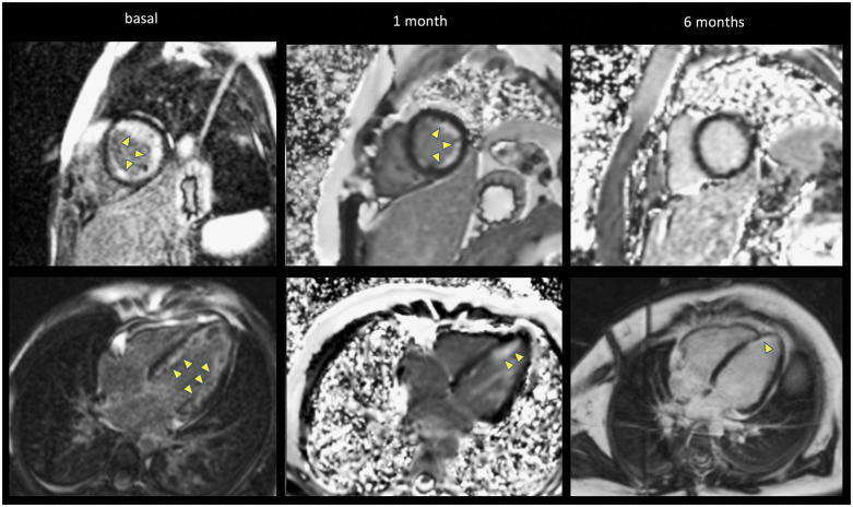
Figure 4.
Late gadolinium enhancement by cardiac magnetic resonance imaging showing the progression of diffuse sub-endocardial enhancement (arrows) of the left ventricular (four-chamber and short-axis views). A large sub-endocardial circumferential signal hyperenhancement is present at the time of diagnosis, decreases at 1 month and resolve at 6 months. Note the different types of magnetic resonance acquisitions present in this figure (2D-IR-FE in the left column, 2D-PSIR-FE in the middle column, and upper right figure and 3D-IR-FE) with the characteristic noise in the background of PSIR images.

