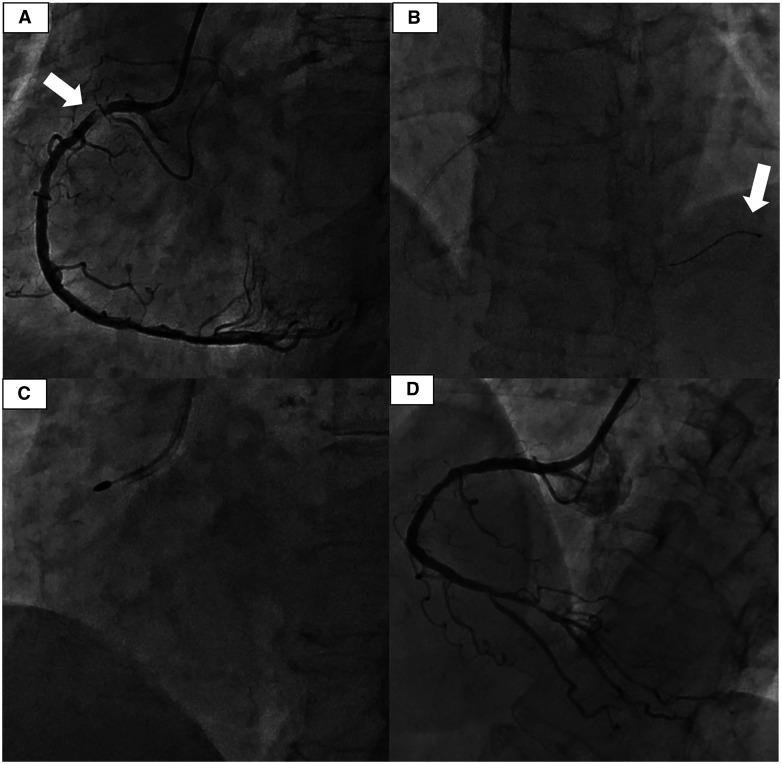
Figure 1.
Result of coronary angiography. (A) Initial angiography. Severely calcified lesion of the proximal right coronary artery (white arrow). (B) The tip of the Rota wire is advanced to the far distal end of the coronary artery (white arrow). (C) Rota burr was used in modifying heavily calcified stenosis. (D) Final PCI results.

