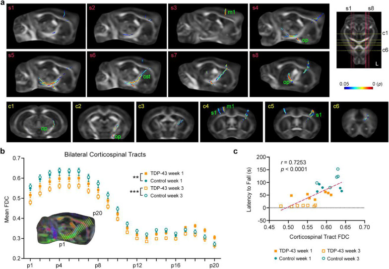
Fig. 3.
DWI reveals progressive microstructural neurodegeneration in TDP-43 mice. a Using connectivity-based fixel enhancement, we compared the change in fibre density and cross-section (FDC) from 1 to 3 weeks after the cessation of Dox feed. When compared to their littermate controls (n = 5), the TDP-43 mice (n = 7) demonstrated significantly reduced FDC in the left corticospinal tract (cst), left optic pathway (op), primary motor cortex (m1) and primary somatosensory cortex (s1). Significant fixels are shown overlaid on the template fractional anisotropy image. The position of sagittal sections (s1–s8, red lines) and coronal sections (c1–c6, yellow lines) are indicated on the axial section at right (‘L’ = left side of the brain). The color bar shows FWE-corrected P-value. b Mean FDC values plotted for each position (from p1 to p20) along the bilateral corticospinal tracts demonstrating progressive damage (i.e., reduced FDC values) to the corticospinal tracts of TDP-43 mice over time. **P < 0.01, ***P < 0.001, FDC values of TDP-43 mice significantly different from control mice. c The caudal corticospinal tract integrity, calculated as the mean FDC value from position p1 to p5, also correlated with motor performance as measured on the rotarod. n = 7 TDP mice, n = 5 control mice

