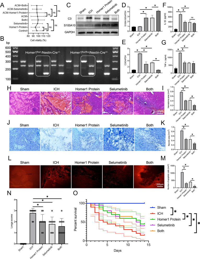
Fig. 7.
Homer1 protein and Selumetinib improve the pathological indexes and prognosis of ICH. A Effects of Homer1 protein and Selumetinib on cell activity [normal medium: F (3, 8) = 0.3731 P = 0.7748; ACM medium: F (3, 8) = 0.5067, P = 0.6885]. B Genotype identification of Homer1flox/flox/Nestin-Cre+/− mice. C WB was used to detect the translation levels of C3 and S100A10 in the brain tissue of mice in each group on the 3rd day after ICH and the results were quantified in D [F (4, 20) = 72.42, P < 0.0001] and E [F (4, 20) = 183.6, P < 0.0001]. The blots are representative of other replicates in those groups. F expression of IL-1β [F (4, 20) = 99.59, P < 0.0001]. G expression of TNF-α [F (4, 20) = 98.31, P < 0.0001]. H Representative photographs of HE staining of brain tissue in each group. I Quantification of result in panel H [F (4, 20) = 279, P < 0.0001]. J Representative photographs of Nissl staining of brain tissue in each group. K Quantification of result in panel J [F (4, 20) = 196.6, P < 0.0001]. L Representative photographs of TUNEL staining of brain tissue in each group. M Quantification of result in panel L [F (4, 20) = 159.6, P < 0.0001]. N Longa scores of different groups on the 3rd day after ICH [F (4, 45) = 34.68, P < 0.0001]. O Survival curve of mice in each group (n = 20) after ICH operation [Log-rank (Mantel–Cox) test: Chi-square = 21.4; df = 4; P = 0.0003]. The data were analyzed using one-way analysis of variance and all data are expressed as the mean ± standard deviation. *P < 0.05 represents a statistically significant difference between the two groups. ns no statistical difference

