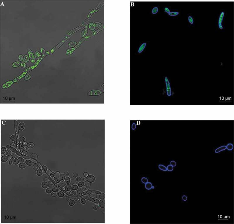
Figure 1.

Incorporation of rhodamine (green) and cell wall labeling with calcofluor white (blue). The images show the overlapping of bright-field and rhodamine fluorescence in the CaS (A) and CaR (C) strains and overlapping fluorescence images of rhodamine and calcofluor white in the CaS (B) and CaR (D) strains
