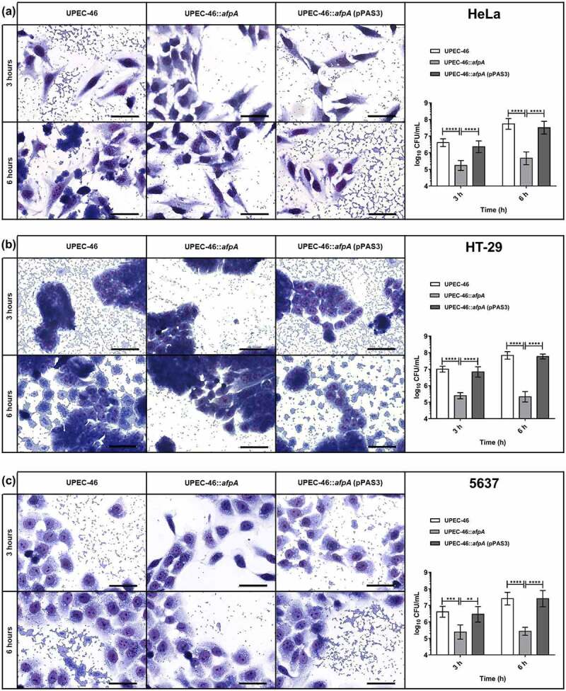
Figure 9.

Adherence assays of UPEC-46 and derivatives with different epithelial cell lineages. The adherence ability was identified after 3 h and 6 h of infection, in the presence of 1% D-mannose, using (a) HeLa (human cervical adenocarcinoma), (b) HT-29 (human colon adenocarcinoma), and (c) 5637 (human urinary bladder carcinoma) cells. In qualitative adherence assays, the evaluation of the AA pattern was performed using light microscopy. Bars = 50 µm. For quantitative adherence assays, the number of cell-adhering bacteria was quantified 3 h and 6 h post-infection as described in materials and methods. The adherence assays with UPEC-46, UPEC-46::afpA, and UPEC-46::afpA (pPAS3) were performed in duplicate and repeated three times. The data presented represent of the mean ± standard deviation. The one-way analysis of variance (ANOVA) followed by Tukey’s multiple-comparison test was used for the statistical analysis. P-value: ** P < 0.01; *** P < 0.001; **** P < 0.0001
