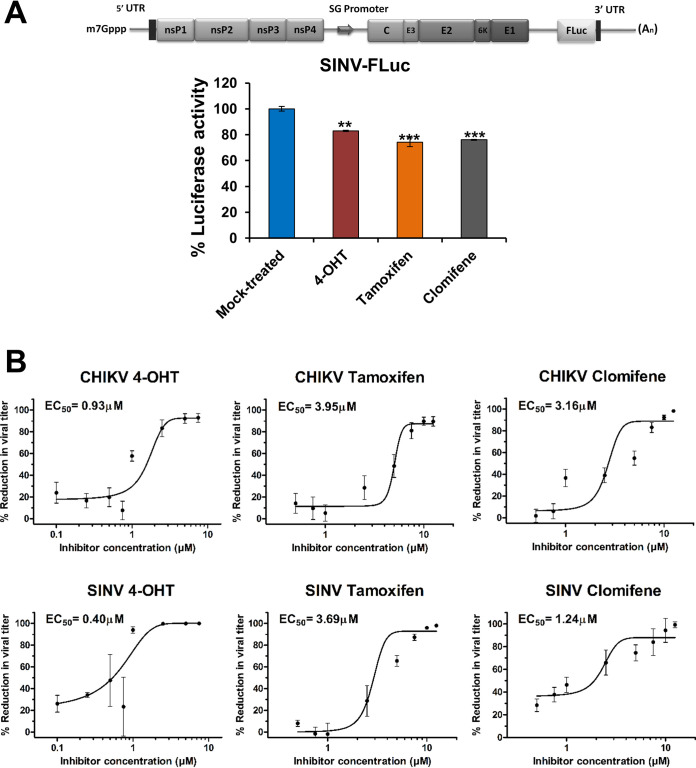
FIG 1.
Primary evaluation of antialphaviral activity of SERMs. (A) Schematic diagram of SINV-FLuc (top) wherein FLuc is placed downstream of an internal ribosomal entry site (IRES). BHK-21 cells were infected with SINV-FLuc at an MOI of 0.1 and treated with SERM compounds or 0.1% DMSO. Luciferase activity of the cell lysate was measured 12 hpi using a firefly luciferase assay kit. Values are the means, and error bars represent standard deviation (n = 3). Statistical significance of the difference in luciferase activity levels between treated and vehicle control-treated cells was assessed by a one-way analysis of variance (ANOVA) test and Dunnett’s posttest; ***, P < 0.001; **, P < 0.01. SG, subgenomic. (B) Plaque assay reveals changes in CHIKV (top) or SINV (bottom) titer on the treatment of virus-infected cells with an increasing concentration of 4-OHT, tamoxifen, and clomifene. The viral titer in the culture supernatant was determined at 24 hpi. The viral titer in corresponding vehicle-treated cells was considered 100%. Data were normalized using GraphPad’s nonlinear regression curve fit, and the calculated 50% effective concentration (EC50) values are summarized in Table 1. Values are the means from three independent experiments, and error bars represent the standard deviation.

