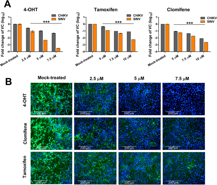
FIG 2.
Antiviral activity of SERMs against CHIKV and SINV in cell culture. (A) Bar plots showing fold reduction in the abundance of vRNA in CHIKV- or SINV-infected Vero cells treated with SERMs compared to vehicle control (VC). At 24 hpi, RNA was isolated and analyzed by qRT-PCR for the expression of the E1 region of the viral genome. Statistical significance of the difference between intracellular vRNA in treated cells and vehicle control-treated cells was assessed by a one-way ANOVA test and Dunnett’s posttest; ***, P < 0.001. Values represent the mean, and error bars are the standard deviation (n = 3). (B) Immunofluorescence microscopy of CHIKV infection in Vero cells upon treatment with SERMs. Virus infection and treatment with SERMs was performed as mentioned in the supporting information (https://www.biorxiv.org/content/10.1101/2021.08.19.457046v2.supplementary-material). At 36 hpi, the infected cells were fixed and stained using an antialphavirus primary antibody and fluorescein isothiocyanate (FITC)-conjugated secondary antibody. 4′,6-Diamidino-2-phenylindole (DAPI) was used for counterstaining cell nuclei. The image represents three independent biological replicates; scale bars, 200 μm.

