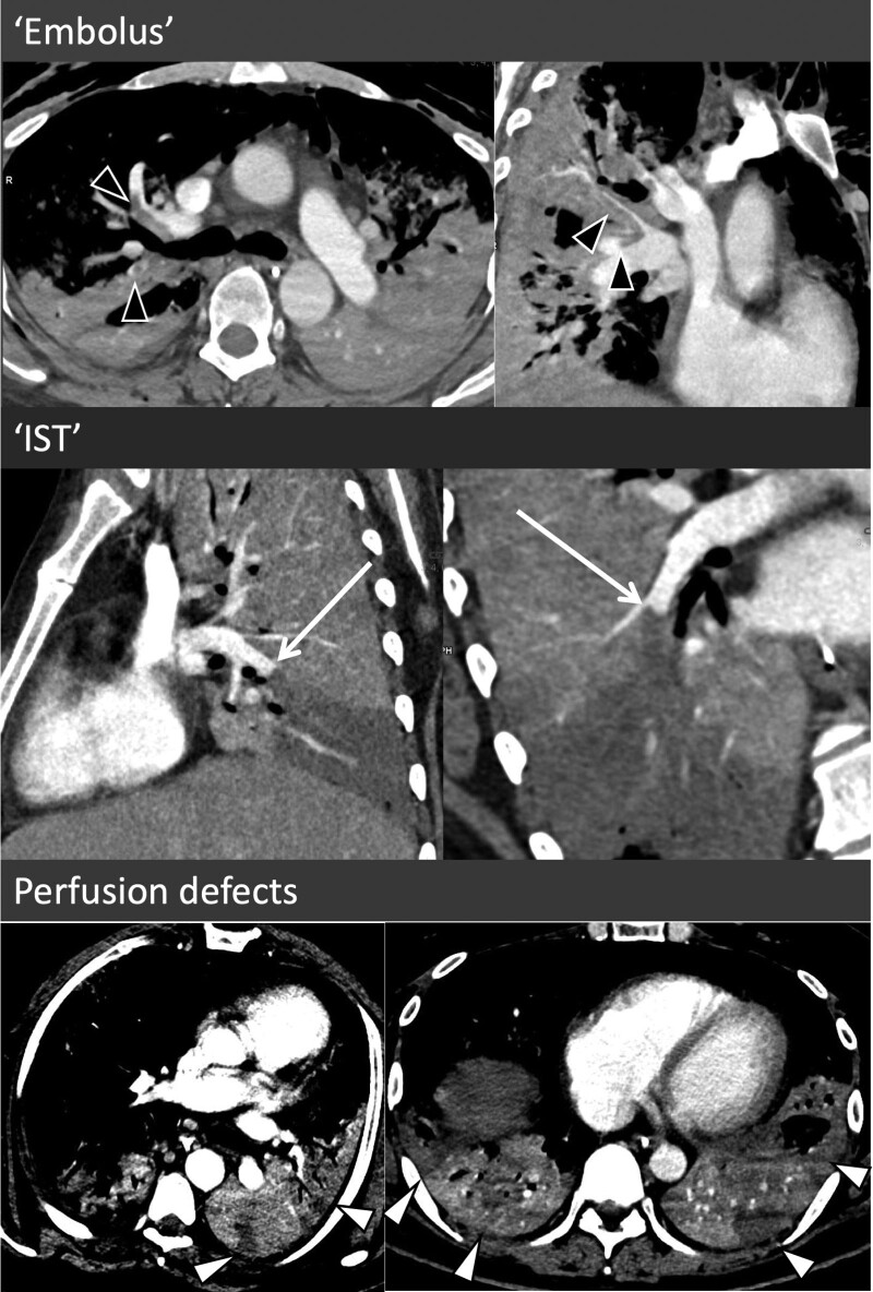
Figure 1.
Morphologic features of pulmonary artery thrombus (PAT) and parenchymal perfusion defects. In the top two images, the PAT exhibits typical characteristic of multiple emboli with central filling defects with peripheral contrast opacification, extending into both branches at a bifurcation points with some of these exhibiting opacification of the pulmonary arteries distally (black arrowheads). In the middle two images, there is complete occlusion of the right lower lobe pulmonary artery with a large wedge perfusion defect in an area of severely disease lung. Rather than the classical crescent of an embolus, the clot is creeping along the outer edge of the vessel toward a more proximal branch (white arrow). The bottom images demonstrate two examples of parenchymal perfusion defects. IST = in situ thrombus.

