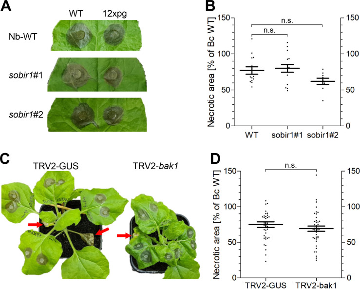
Fig 7. Infection of Nicotiana benthamiana WT and sobir1 mutants, and plants silenced for bak1, by B. cinerea WT and 12xpg mutant.
A: Lesion formation on N. benthamiana WT and sobir1 leaves (48 h.p.i.). B: Necrosis induction of 12xpg mutant relative to WT. B. cinerea. WT-induced lesions (on N.b. WT: 143.8±9.2; on N.b. sobir1#1: 129.5±10.9; on N.b. sobir1#2: 145.1±17.2 mm2) and relative lesion sizes of 12xpg were not significantly different (n.s.) between N.b. WT and sobir1 mutants (one way ANOVA and Dunnet’s post-hoc test). Data are from three independent experiments, with two plants and two or three leaves per plant each. C: Lesion formation by B. cinerea WT (upper left sides of leaves) and 12xpg mutant (upper right sides) on plants subjected to VIGS (48 h.p.i.). Sites of TRV2 inoculation for VIGS induction are indicated by red arrows. Note the stunted growth and wrinkled leaves of the plant silenced with TRV2-bak1. D: Necrosis induction of 12xpg mutant relative to WT. WT-induced lesions (on N.b. TRV2-GUS: 191,5±9.2; on N.b. TRV2-bak1: 178.4±9.2 mm2) and relative lesion sizes of 12xpg were not significantly different (n.s.) on these plants (one way ANOVA and Tukey’s post-hoc test). Data are from four independent experiments with two batches of VIGS-silenced leaves, with two plants per experiment and two or three leaves per plant.

