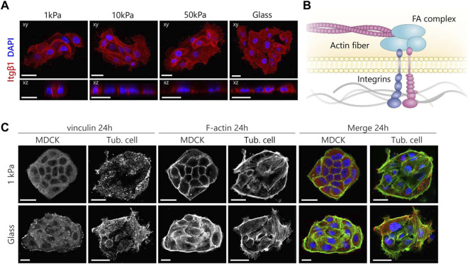
FIGURE 4.
MDCKs and tubuloid-derived cells show a different organization of the mechanosensing machinery according to substrate stiffness. (A) Representative fluorescent images of β1 integrin expression in tubuloid-derived cells cultured on substrates for 24 h with variable elastic moduli. Cells were stained for β1 integrin (red) and nuclei (blue). (B) A graphical illustration displaying the mechanotransduction pathway that acts via the dynamic link the FA complex forms between integrins and the actin cytoskeleton. (C) Immunofluorescence images displaying the differences in maturation and organization of the focal adhesions in red (vinculin) and the cytoskeleton in green (F-actin) after 24 h. Scale bars are 20 µm.

