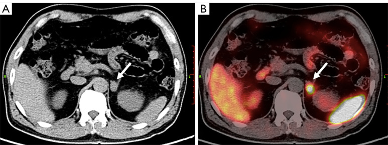
Figure 3.
PCC in a 66-year-old male. (A) CT image showing a hypointense tumor at the left adrenal gland (arrow). Biochemical tests of functional adrenal tumors including PPGLs were all negative. (B) This tumor showed intense uptake of 68Ga-DOTAT-TATE in the CT and PET fusion image (arrow). The SUVmax was 19.1, SUVR was 2.2, score of KS was 3 respectively. PCC was diagnosed before surgery, and was later confirmed by histologic results. PCC, pheochromocytoma; CT, computed tomography; PPGLs, pheochromocytomas/paragangliomas; 68Ga-DOTA-TATE, 68Ga-DOTA(0)-Tyr(3)-octreotate; PET, positron emission tomography; SUVmax, maximal standardized uptake value; SUVR, ratio between the SUVmax of the lesion and the SUVmean of the liver; SUVmean, mean standardized uptake value; KS, Krenning scale.

