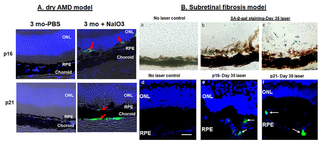
Fig.7. Evidence for senescence in RPE cells in dry AMD (A) and subretinal fibrosis (B) mouse models.

Dry AMD refers to confocal images on day 7 after mice were given NaIO3 (20mg/kg BW) intravenously as a single injection and subretinal fibrosis was produced from laser-irradiation on Day 35. Evidence for the presence of senescent RPE cells by SA-β-gal, p16, and p21 expression in day 35 post-laser retina from 8 wk old mice. Panels (b,c) show SA-β-gal + cells (arrows; blue-green cells). Panels (e,f) show positive immunoreactivity for p16 and p21 (red arrows in A and white arrows in B). No primary antibody control for p21 and p16 was used as a negative control (not shown). Scale bar 5μm. RPE- Retinal pigment epithelium, ONL- Outer nuclear layer.
