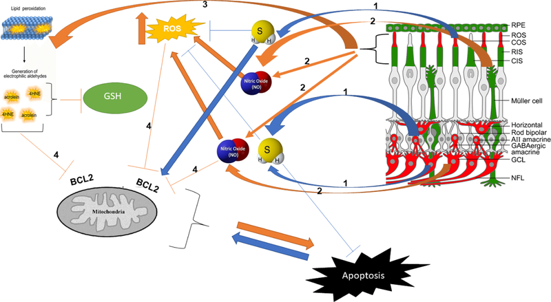
Figure 1.
Lipid peroxidation, NO, and molecules produced by TSP enzymes affect the apoptosis pathway in retinal degenerative diseases. Schematic representation of H2S and NO molecules produced in retina and interaction with ROS products. Key molecules and components of the pathway: (1) CBS and CSE are the main enzymes of TSP involved in H2S and GSH production in the retina. Physiological level of H2S is shown to have neuroprotective effects. (2) NO is a gaseous molecule that affects BCL2 and mitochondrial function, and excessive NO produced in retinal degeneration is detrimental to retinal cells. (3) The photoreceptor membrane is rich in PUFA and is vulnerable to peroxidation because of chronic light exposure. High metabolic rate and high levels of oxygen levels result in ROS production. Increased levels of ROS lead to the peroxidation of PUFAs. (4) Acrolein and 4HNE are the metabolites of PUFAs peroxidation that, along with ROS, can affect GSH and BCL2 and cause mitochondrial dysfunction. The schematic of the retina on the top right was modified from Figure 9 of Badiei et al. Exp. Eye. Res. (2019) and shows immunolocalization of CBS (red) and CSE (green) in retinal neurons and subcellular compartments.

