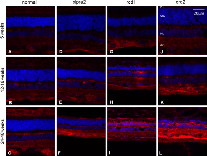
Figure 3.
Immunohistochemistry of CBS (red fluorescence) in normal and mutant retinas of different ages. With the progression of the disease, immunolabeling intensity increases in the GCL. Labeling of the inner and outer plexiform layers also increases in the older rcd1 and crd2 mutant retinas. DAPI (blue) nuclear stain. Scale bar: 20 μm; PR: Photoreceptor cells, ONL: outer nuclear layer; INL: inner nuclear layer; GCL: ganglion cell layer.

