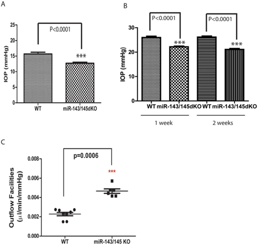
Fig. 2. Reduced IOP and increased outflow facilities in miR-143/145 dKO mice.
(A) Reduced intraocular pressure (IOP) in miR-143/145 dKO mice. ***p < 0.0001; (B) Reduced intraocular pressure in miR-143/145 dKO mice in an experimental glaucoma model. The IOP of the mice was measured at 1 and 2 weeks after microbeads injection into the eye. ***p < 0.0001; (C) Significant increase in outflow facilities in 16-month-old miR-143/145 dKO mice compared to that in WT control mice. ***p < 0.0006. Figure adapted from Li et al. (2017) with permission.

