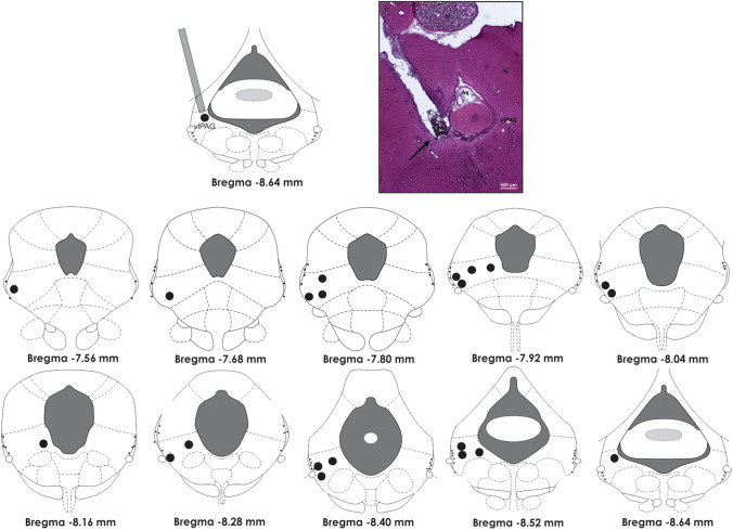
Fig. 1.
Schematic view (in the top left corner) of a guide-cannula inserted in the ventrolateral column of the periaqueductal gray matter (vlPAG). A photomicrograph of a representative site (black arrow) of microinjections of drugs in vlPAG is provided in the top right corner. In the bottom, drawings of midbrain transverse sections across rostrocaudal extensions of the periaqueductal gray matter, depicting the representation of the histologically confirmed injection sites (black circles) of either physiological saline or NMDA in the vlPAG. The number of points in the figure is fewer than the total number of rats because of overlapping injection sites. vlPAG ventrolateral columns of the periaqueductal gray matter, Pi pineal gland, Aq aquaeductus Sylvii, IC inferior colliculus. Calibration bar: 500 µm

