
Figure 4.
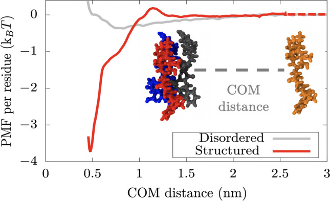
Atomistic potential-of-mean-force (PMF) dissociation curve of an 8-amino acid segment (PDB code: 6BZM) of NUP-98 protein from a -sheet structure of 4 peptides (of the same sequence) as a function of the center of mass distance (COM) using the a99SB-disp force field116. Red curve depicts the interaction strength among peptides with a well-defined folded structure, kinked -sheet structure, while grey curve represents the interaction strength among the same segments but when they are disordered. The binding interaction strength difference between disordered and ordered peptides differs by almost an order of magnitude. The same calculations performed using the CHARM36m force field117 are shown in Fig. S9, where the obtained difference in binding strength between disordered and structured peptides is of the same order.