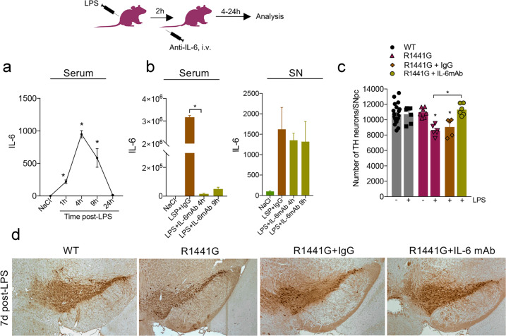
Fig. 4. Inhibition of peripheral IL-6 rescues LPS-induced SNpc cell loss in R1441G mice.
a Time-course of serum IL-6 secretion in R1441G mice at different time points post-LPS. Data are mean ± SEM, n = 5–8. *p < 0.05. b Anti-IL-6 antibody significantly inhibits LPS-induced IL-6 up-regulation in serum, with no change was seen in the SN. Data are mean ± SEM, n = 3–5. *p < 0.05. For each group, data in LPS-treated groups are expressed as the percentage of NaCl control. c Neutralization of peripheral IL-6 completely rescues LPS-induced SNpc DA neuron in R1441G mice. Data are mean ± SEM, n ≥ 4. *p < 0.05 vs WT or vs group indicated on the graph. d Representative images of TH-positive DA neurons in the SNpc of LPS-treated WT, R1441G, and R1441G that received either anti-IL-6 mAb or IgG. Sections are matched at the same level of the SN (Bregma −3.08–3.16 mm) (Paxinos and Franklin, 2001). Scale bar = 100 μm.

