Figure 7.
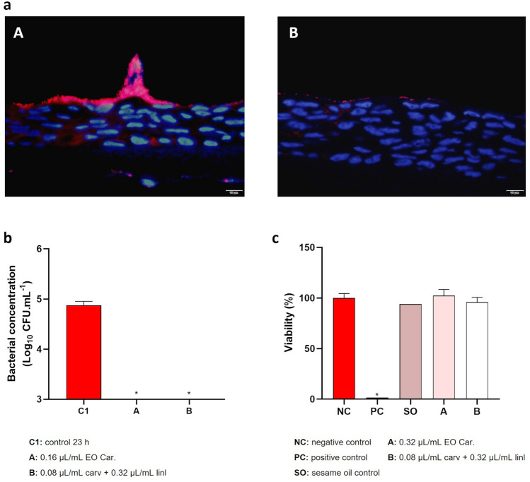
Colonization of reconstituted human vaginal epithelium and cytotoxicity of T. capitata EO. (a) Microscopic images of reconstituted human vaginal epithelium after 23 h of colonization with G. vaginalis UM137 (A) and after 9 h of G. vaginalis UM137 colonization with 14 h of contact with EO Car. (B). (b) Results of cells culturability after colonization of vaginal epithelium. Results are represented as mean of Log (CFU.mL−1) with a limit of detection of Log = 3. Error bars represent s.d. Statistical analysis was performed using t-test (n = 3 for control and EO assay; n = 2 for carvacrol + linalool assay). Differences are represented by * when comparing with control 23 h, when p < 0.05. (c) Results of cytotoxicity of EO and combination carvacrol + linalool on the vaginal tissue are the mean of percentage (%) of viability. Error bars represent s.d. Statistical analysis was performed using a t-test (n = 4 for EO assay; n = 3 for controls and carvacrol + linalool assay). Differences are represented by * when comparing with the negative control. Statistical analysis did not include sesame oil control (n = 1).

