Fig. 3.
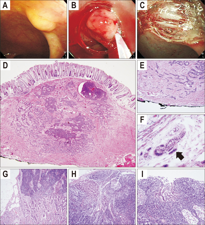
Case of small rectal neuroendocrine tumor with multiple lymph node metastases. Endoscopic (A-C) and histopathologic findings of rectal neuroendocrine tumor (D-F) and perirectal lymph nodes (G-I). A small, yellowish subepithelial tumor was located at the lower rectum (A). The lesion was completely removed by endoscopic mucosal resection (B, C). Microscopic findings showed monotonous small round cells arranged in a solid and pseudoglandular pattern (D, H&E, ×1; E, H&E, ×60). Angiolymphatic invasion was observed with monotonous small cell clusters (black arrow) (F, H&E, ×200). Monotonous cell clusters were also observed in three perirectal lymph nodes (G-I, H&E, ×200).

