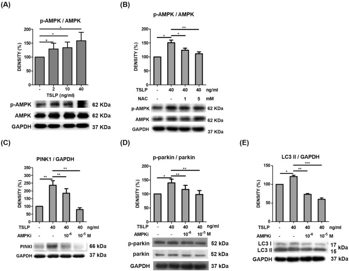
Fig. 6.
AMPK activation promoted TSLP-induced mitophagy-related protein expression. A TSLP (2, 10 and 40 ng/mL) significantly increased phospho-AMPK (p-AMPK) signal intensity, as shown by western blot analysis. B TSLP-induced p-AMPK signal intensity was suppressed by the antioxidant N-acetylcysteine (NAC). TSLP-induced protein signal intensities of C PINK1, D phospho-Parkin (p-Parkin), and E LC3 were suppressed by an AMPK inhibitor, as shown by western blot analysis. The standard deviations of the optical density data were calculated from three independent experiments, and one representative result of the set of three is shown. *p < 0.05, **p < 0.01 and ***p < 0.001

