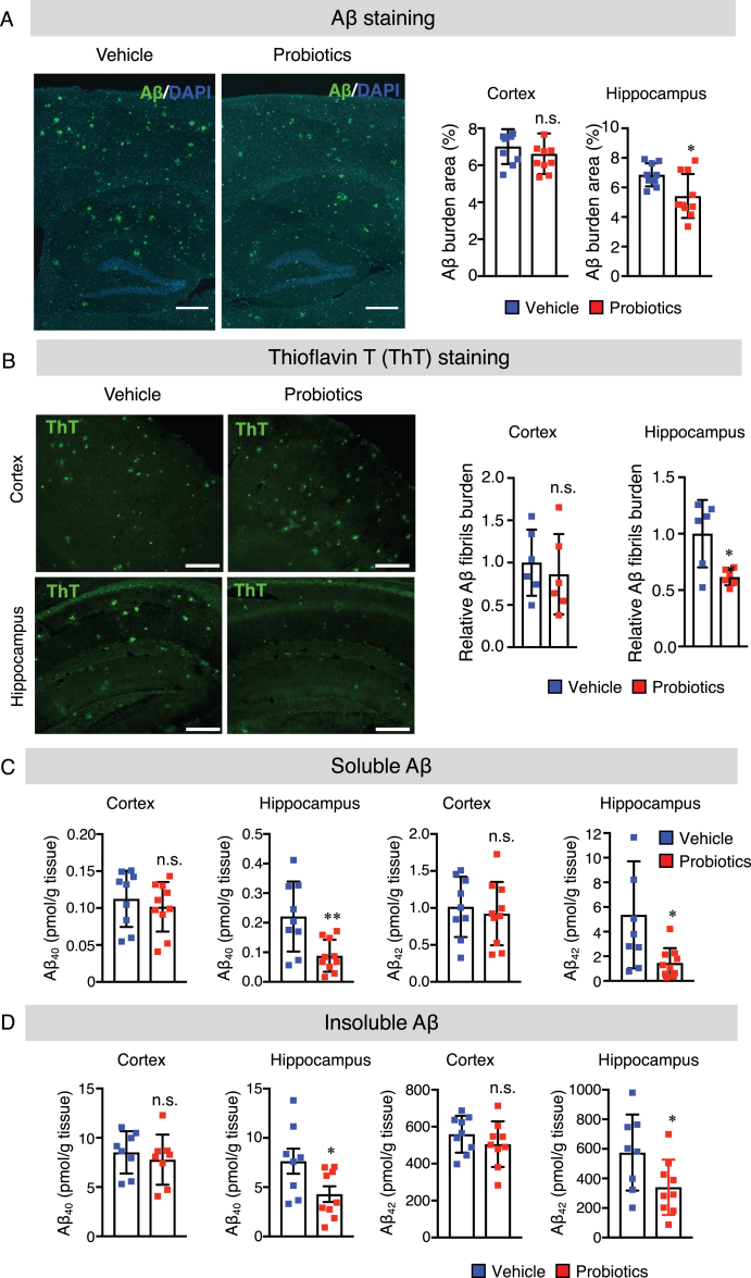
Fig. 2.
B. breve MCC1274 supplementation reduces Aβ plaque burden, Aβ levels, and Aβ fibrils in the hippocampus of AppNL-G-F mice. A) The representative fluorescent images of Aβ plaque burden detected by anti-Aβ antibody (82E1), which recognizes both Aβ40 and Aβ42 (left panel). Aβ burden areas including the cortex and hippocampus were quantified as the percentage of immunostained area divided by all cortical and hippocampal areas (right panel). Scale bars are 250μm. B) The representative fluorescent images of Aβ fibril were detected by thioflavin T (left panel). Relative Aβ fibril burden in both cortex and hippocampus were quantified (right panel), Scale bars are 100μm. Sandwich ELISA result of cortical and hippocampal levels of soluble Aβ40 and Aβ42 (C) and insoluble Aβ40 and Aβ42 (D) of AppNL-G-F mice. Aβ levels were normalized to each tissue weight. Data are expressed as the mean±SD, n = 9-10, *p < 0.05, **p < 0.01 compared with the vehicle group, as determined by Student’s t-test.

