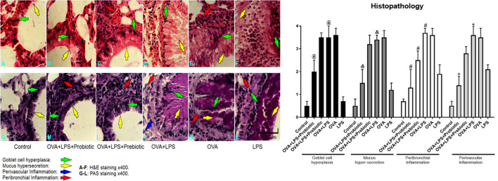
Fig. 4.
Lung histopathology. Lung sections were stained with H&E and PAS and the infiltration of inflammatory cells (eosinophils) into perivascular and peribronchial, along with goblet cell hyperplasia, and mucus hypersecretion were studied. The histopathological results showed that infiltration of inflammatory cells into the perivascular and peribronchial regions, goblet cell hyperplasia and mucus hyper-production were increased significantly (p < 0.05) in OVA-LPS-induced asthmatic mice compared with the PBS sensitized mice. A result analysis revealed a significant (p < 0.05) reduction in perivascular and peribronchial inflammation, goblet cell hyperplasia and mucus hyper-production in the probiotic-treated groups compared with the OVA-LPS group. Treatment with prebiotics could significantly (p < 0.05) control only peribronchial inflammation. The significant difference of the results (p < 0.05) between treated groups and non-treated OVA-LPS groups was shown with symbols that include: * for perivascular inflammation, # for peribronchial inflammation, & for mucus hypersecretion, and @ for goblet cell hyperplasia. In the histopathologic sections, hyperplasia of the goblet cells were shown by green arrow, hypersecretion of mucus was shown by yellow arrow, perivascular eosinophilc inflammation was shown by blue arrow and peribronchial eosinophilc inflammation was shown by red arrow

