Fig. 1.

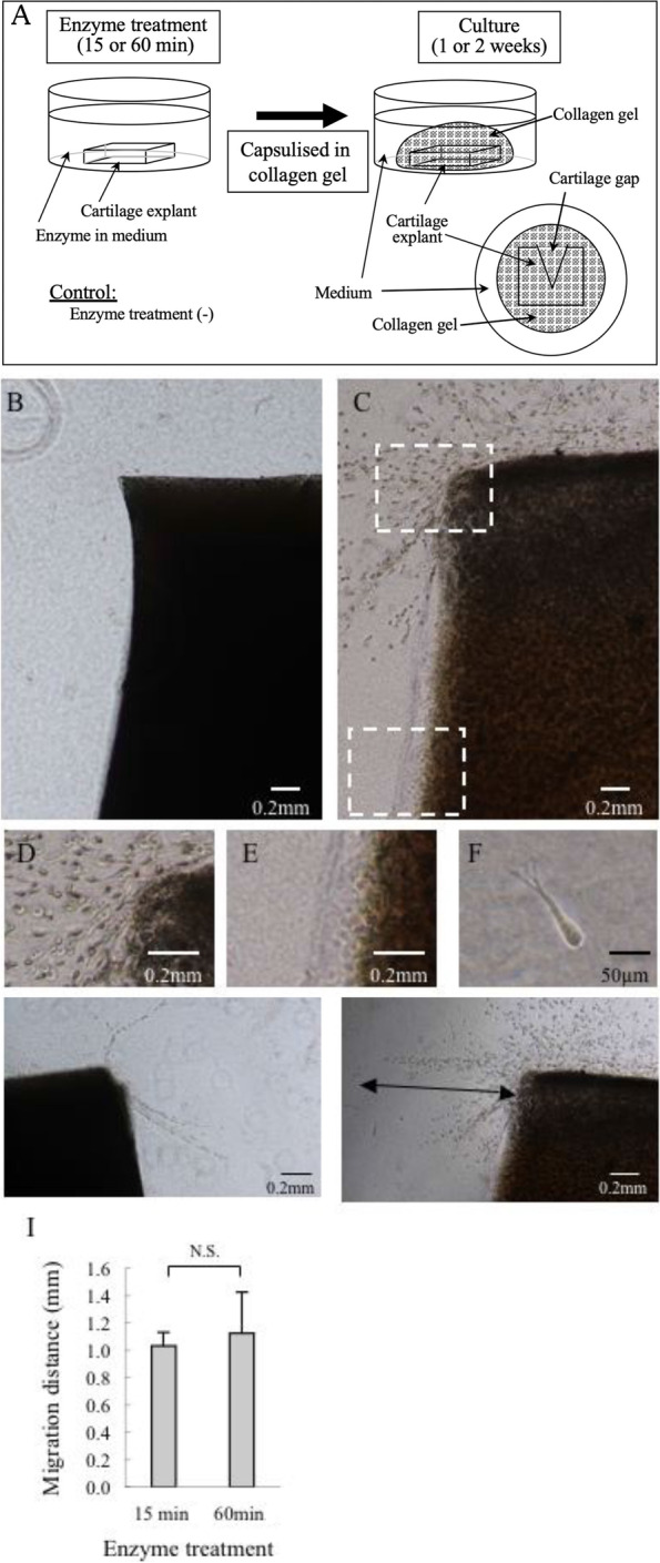
Culture of full-thickness cartilage explants and analysis of the effects of enzymatic treatment on chondrocyte migration. Cartilage explants harvested from the patellar articular cartilage of a 3-month-old pig were enzymatically treated (0.2% actinase and 0.02% collagenase P) in culture medium (Dulbecco’s modified essential medium supplemented with 10% fetal bovine serum, 0.2% ascorbic acid, and 0.5% antibiotics) for 15 or 60 min, encapsulated in type-I collagen gel, and cultured for 1 or 2 weeks (A). Cell migration was examined using a phase contrast microscope. No migrating chondrocytes were found without enzymatic treatment (B). Abundant cells migrated from the superficial zone of a cartilage explant treated with actinase and collagenase (C). Hyper magnification of the inset in C showing abundant cells migrating from the superficial zone (D) and no cells migrating from the deep zone (E). Hyper magnification of a representative migrating cell showing spindle shape with pseudopodia (F). Migration distance and the number of migrating cells were compared between the 15-min treatment group (G) and the 60-min treatment group (H). Arrow indicates migratory distance. Migratory distance was compared between the two groups (I). N.S.: not significant (The Mann-Whitney U test)
