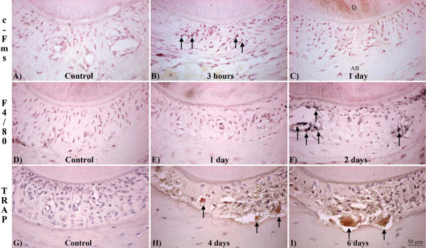
Figure 2.
Expression of OC differentiation markers in the mesial (compression) side of the PDL surrounding the distal-palatal root of the maxillary first molar during OTM. (A,B,C) c-Fms–positive cells (arrows). (A) Control. (B) 3 Hours. (C) 1 Day of force. (D,E,F) F4/80-positive cells (arrowheads). (D) Control. (E) 1 Day. (F) 2 Days of force. (G,H,I) TRAP-positive cells (red). (G) Control. (H) 4 Days of force. (I) 6 Days of force. (A,B,C,D,E,F) Counterstained with neutral red. (G,H,I) Hematoxylin and eosin (H&E). D indicates dentin; AB, alveolar bone.

