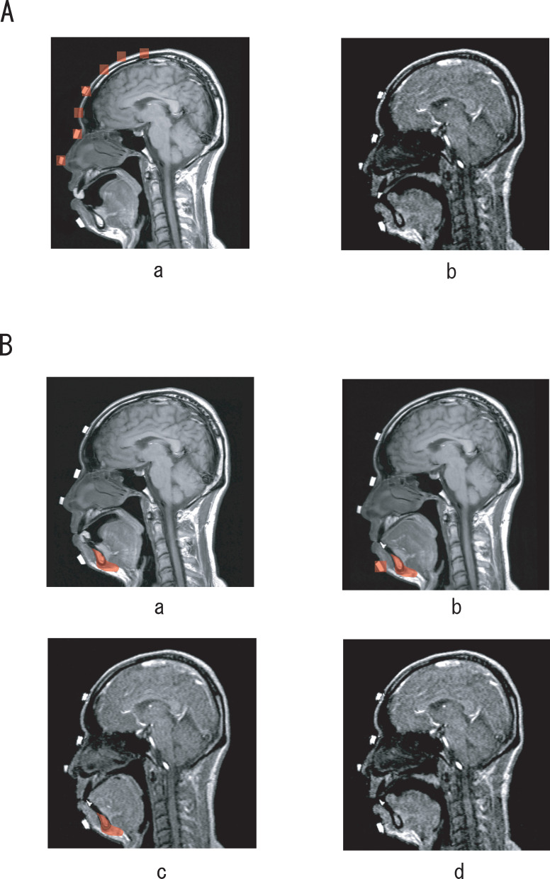
Figure 3.
(A) Superimposition of the upper incisor and hard palate. Landmarks on the nasal tip, nasal root, forehead, and points along the cranium (Aa) were used for superimposition. (Ab) One of the MRI movie images obtained from this superimposition. (B) Superimposition of the lower incisor. First, the landmark at the symphysis (Ba) was used to superimpose the lower incisor onto the image taken at rest with a TSE-T1 sequence. The landmarks on the chin and symphysis (Bb) were then used to superimpose the lower incisor onto the image taken at rest with a GRE sequence. Finally, the landmark at the symphysis (Bc) was used to superimpose the lower incisor onto an MRI movie image. (Bd) The image obtained after superimposition.

