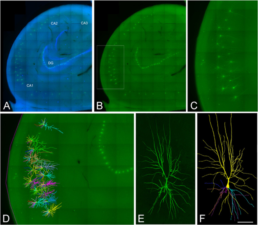
Figure 1.
Confocal microscopy images of human neurons injected with Lucifer yellow in the hippocampus. A, B, Labeled pyramidal cells (green) and DAPI staining (blue) in different regions of the human hippocampus, including CA1, CA2, CA3, and the dentate gyrus region (DG). C, Higher magnification image of the boxed region shown in B. D, 3D reconstructed cells superimposed on the confocal image shown in C. E, F, High-magnification image z projection showing an injected CA1 pyramidal cell (E) and the 3D reconstruction of the same cell (F). Scale bar: 1100 μm (A, B), 460 μm (C, D), and 100 μm (E, F). Image taken from Benavides-Piccione et al. (2020).

