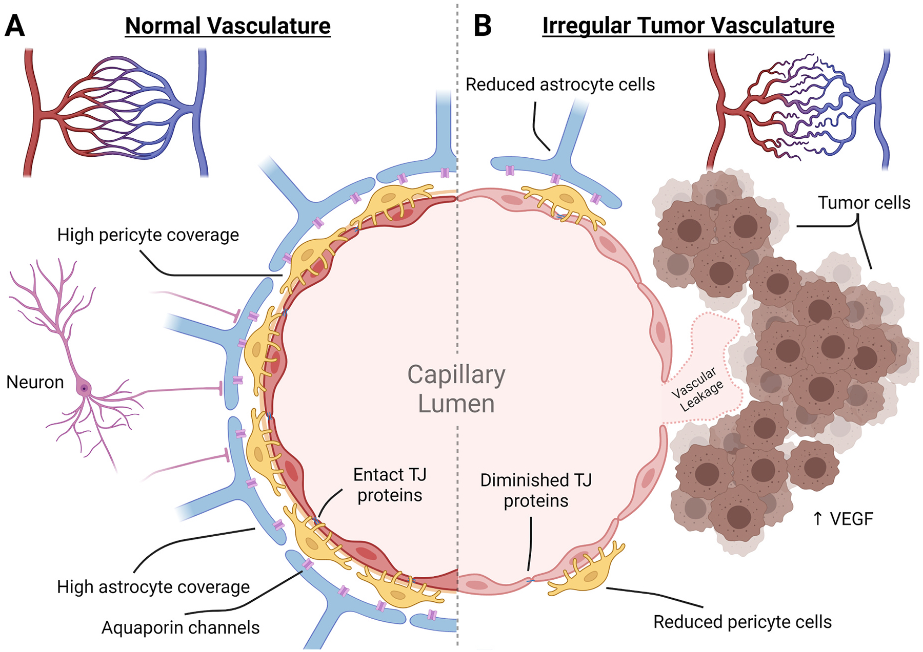
Fig. 1. Comparison of the neurovascular unit of the blood brain barrier and blood tumor barrier.

Normal vasculature of the brain develops from pial arteries to arterioles to capillaries where transfer of nutrients and oxygen can take place. In the normal tissue, capillaries are spread out somewhat uniformly to ensure equal distribution. In the BTB, there is a reduced presence of capillary endings and an irregular overlay creating areas of hypoxic tumor tissue. (A) In the normal tissue capillaries, the neurovascular unit controls the influx of materials by forming a non-fenestrated endothelial cell layer that is tightly bound together with TJ proteins. These are enveloped by pericyte cells and astrocytic endfeet that aid in maintaining the endothelial cell transport proteins, TJ proteins, induction of angiogenesis, and influx of ions and fluids. This is controlled by local input as well as neuronal demand that can interact directly or through astrocytic channels. (B) Within the BTB, integrity is compromised. Tumor cells attach to the outer vascular wall and induce vascular growth through VEGF expression. The new capillary bed is irregular and leaves areas of the tumor hypoxic. The new vasculature becomes the foundation of the BTB and has been found to have reductions in astrocytic endfeet coverage, pericyte cells, TJ proteins, and neuron attachment. The resulting capillary wall of the BTB is susceptible to neurotoxic leakage.
