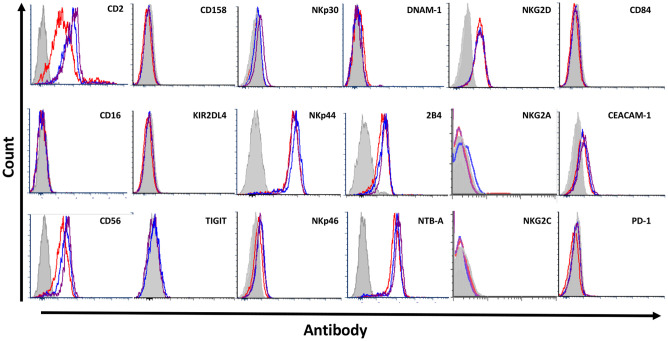
Fig 2. Characterization of surface receptor expression.
1 out of 4 representative FACS stainings against various inhibitory and activating NK cell markers (CD2, CD56), KIR (CD158, KIR2DL4), activating receptors (NKp30, NKp44, NKp46, NKG2D, NKG2C, DNAM-1, 2B4, NTB-A, CD84), and inhibitory receptors (TIGIT, CEACAM-1, PD-1, NKG2A) grey = background staining, red = NK-92med., blue = RPMIf, violet = RPMIp.

