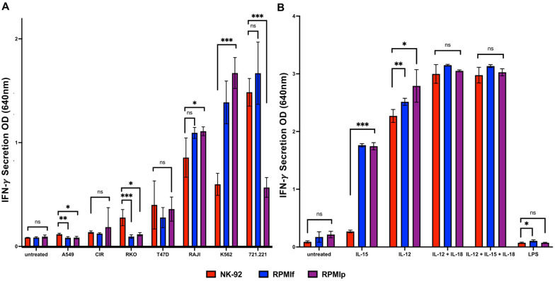
Fig 3. Comparison of secretory potential after activation of NK-92 cells in different growth conditions.
A) + B) Secretion of IFN- γ; A) Secretion after activation with tumor cells for 48h at 37°C (721.221, A549, C1R, K562, RAJI, RKO, T47D), B) Secretion after activation with interleukins after 48h of incubation at 37°C (IL-12, IL-15, IL-12 + IL-18, IL-12 + IL-15 + IL-18, LPS); red = NK-92med., blue = RPMIf, violet = RPMIp. *p<0.05, **p<0.005, ***p<0.001 compared between the NK-92 in NK-92 medium as control and the NK-92 in RPMI + IL-2 medium (two-tailed ANOVA test + multiple unpaired T-tests). Shown is 1 representative out of 5 assays performed.

