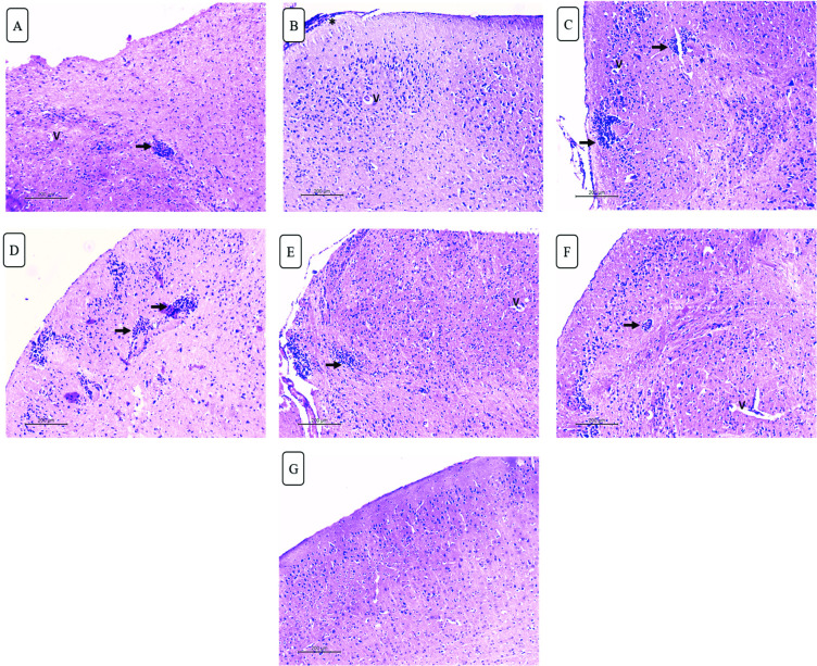
Fig 10. H&E-stained brain sections of different studied groups.
(A) Infected untreated group showing mononuclear parenchymal infiltrate (black arrow) with perivascular oedema (v) (X100). (B) Mild improvement is seen in spiramycin treated group showing residual meningitis (asterisk) and perivascular oedema. (X100). (C-F) Mononuclear parenchymal infiltrate (black arrow) and perivascular oedema (v) are still noted in propolis, CS/Alg NPs, spiramycin CS/Alg NPs and propolis CS/Alg NPs treated groups respectively (X100). (G) Best results are seen in spiramycin/ propolis loaded CS/Alg NPs treated group with normal neurons, no meningitis or mononuclear infiltrate (X100).

