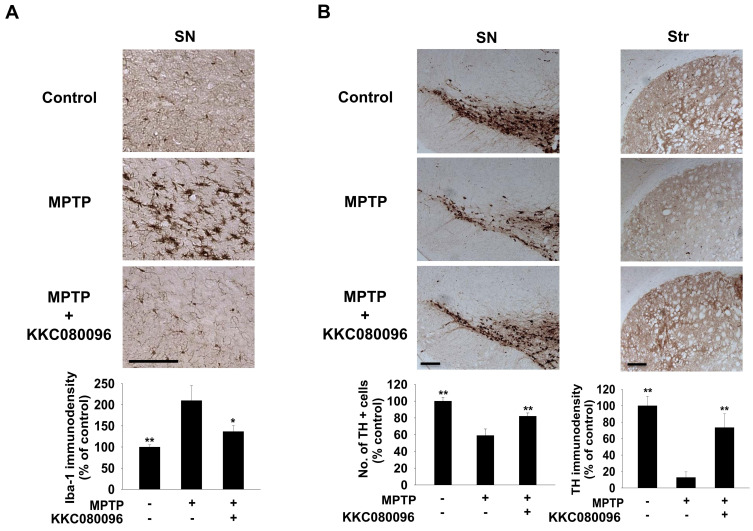
Fig. 3. KKC080096 suppresses microglial activation and protects dopaminergic neurons in MPTP-treated mice.
Immunohistochemistry in mice administered MPTP alone only or co-treated with 30 mg/kg KKC080096. (A) Photomicrographs of the nigral sections showing Iba-1 expression (top) and quantitative analysis of the Iba-1-immunopositive microglia in the SN using densitometry (bottom). Scale bar = 200 μm. (B) Photomicrographs of the nigral (top left) and striatal (Str) (top right) sections immunostained against TH and quantitative analysis of the TH-immunopositive neurons in the SN by counting (bottom left) and TH-immunopositive terminals in the striatum by densitometry (bottom right). Scale bars = 200 μm. The data are expressed as the percentage of vehicle control group ± SEM; *P < 0.05, **P < 0.01 vs MPTP group.

