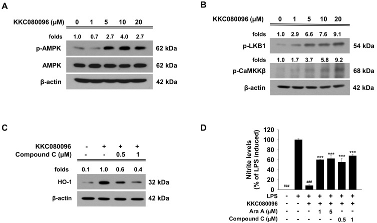
Fig. 5. KKC080096 activates AMPK, LKB1, and CaMKKβ in microglial cells.
BV-2 cells were treated with various concentrations of KKC080096 for 15 min. The cells were harvested and (A) the levels of total and phosphorylated AMPK and (B) phosphorylated LKB1 and CaMKKβ were detected by western blotting with β-actin as a loading control. (C) The cells were pretreated with Compound C for 1 h, treated with KKC080096 (20 μM), and then cultured for additional 24 h. Western blotting was performed against HO-1 using β-actin as loading control. (A-C) The number on each gel pictogram represents the fold change with respect to untreated control (A and B) or KKC080096-treated control, calculated from densitometric value that had been normalized against β-actin. (D) The cells were pretreated with Ara A or Compound C for 1 h, treated with KKC080096 (20 μM) and/or 0.2 μg/ml LPS, and then cultured for additional 24 h. The nitrite level was determined using the Griess assay. The data are expressed as percentage of LPS-induced control ± SEM; ###P < 0.001 vs LPS-treated; ***P < 0.001 vs (KKC080096 + LPS)-treated.

